Blood Vessels
As you'll soon learn, the heart is responsible for sending the various components of blood around the body to take valuable oxygen to cells and to remove waste products such as carbon dioxide back to the lungs to be expelled as we breathe out.
If we regard blood cells and the various components of blood as transport vessels (because in effect, that's what they do, they transport oxygen and carbon dioxide and other substances around the body) we need a suitable network of "motorways" and "main roads" for this to happen. The blood vessels of the cardiovascular system are the motorways and main roads, and these are what we will look at.
There are three main blood vessels to be aware of, arteries, veins and capillaries.
|
|
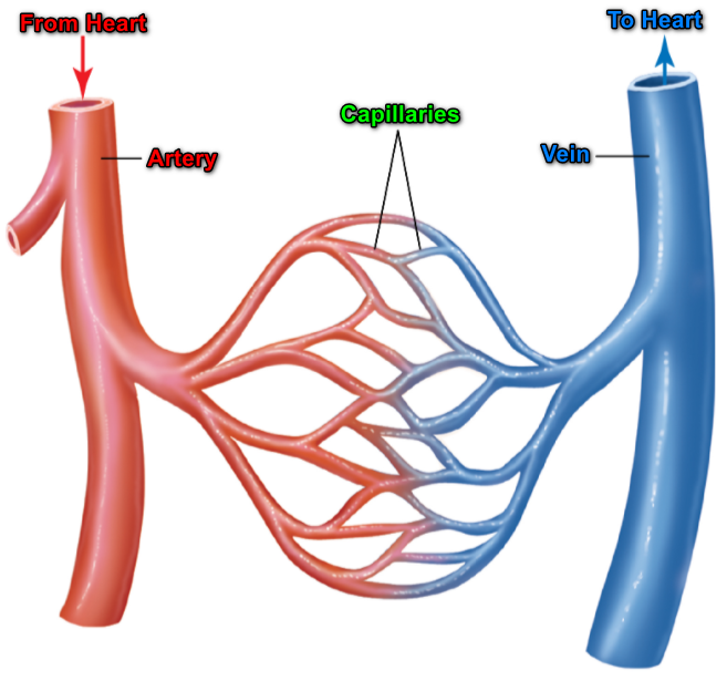
In the image shown, the cross sections of the three blood vessels tell a lot about how they are all adapted to their specific functions. The "tube" that the blood flows along is called the "lumen" and in the case of the artery is quite narrow. The walls of the artery are very thick with muscular tissue so that they can withstand the pressure of the blood being forced out of the left ventricle of the heart to commence its journey around the body. In the case of the vein, the "lumen" is larger and the thickness of the wall of the vein is considerably less than that of the artery because the pressure of blood in the vein is quite a bit lower. Veins have one-way valves at regular intervals along the length so that during beats of the heart, the blood cannot attempt to flow the wrong way. If any of these valves become compromised or damaged and are therefore unable to open and close properly, blood pressure can build up in the blood vessel and cause problems. Sufferers with compromised venous systems of this type usually wear elasticated support garments to assist with circulation. The capillary is considerably smaller and thinner than the other two, the walls of the capillary are usually only one cell thick which allows for efficient gas exchange known as "a short diffusion pathway". |
Arteries transport blood away from the heart, and as we have said already this is done under high pressure so the walls of the artery are quite thick. Arteries eventually produce a network of fine capillaries where the substance exchanges take place. Capillaries subsequently join back up to form veins which are responsible for returning blood to the heart.
Blood flow refers to the movement of blood through a vessel, tissue, or organ, and is usually expressed in terms of volume of blood per unit of time. It is initiated by the contraction of the ventricles of the heart. Ventricular contraction ejects blood into the major arteries, resulting in flow from regions of higher pressure to regions of lower pressure, as blood encounters smaller arteries and arterioles, then capillaries, then the venules and veins of the venous system.
Hydrostatic pressure is the force exerted by a fluid due to gravitational pull, usually against the wall of the container in which it is located.
One form of hydrostatic pressure is blood pressure, the force exerted by blood upon the walls of the blood vessels or the chambers of the heart. Blood pressure may be measured in capillaries and veins, as well as the vessels of the pulmonary circulation; however, the term blood pressure without any specific descriptors typically refers to systemic arterial blood pressure, that is, the pressure of blood flowing in the arteries of the systemic circulation. In clinical practice, this pressure is measured in mm Hg and is usually obtained using the brachial artery of the arm.
You may have heard at some point, someone discussing blood pressure and using an expression of <one number> over <another number> such as "130 over 80".
These numbers refer to the systolic and diastolic blood pressure. When your blood pressure is taken it is measured at two points:
1. When the artery is full of blood immediately after a heart beat
2. When the artery is full of blood but in a relaxed state (between beats)
Immediately after the heartbeat, when the blood pressure in the artery is at its highest is the systolic pressure, and during a "relax" between beats the pressure is recorded as the diastolic.
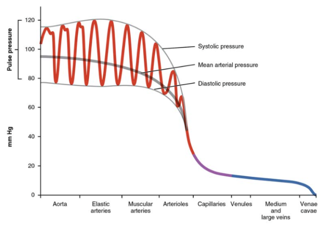
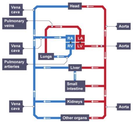
The diagram shows the approximate pressures in millimetres of mercury that you would expect to find in the difference "grades" of blood vessels from the aorta in the arterial system across to the vena cavae in the venous system. If you think about it, it makes sense for blood to leave the heart under high pressure but to return under low pressure and the arterial and venous system blood vessels are designed to accommodate that.
Although the blood pressure from the artery is measured (usually the upper left arm, the brachial artery) as a ratio of the systolic (during beats and diastolic (between beats) average can be recorded which is known as the mean arterial pressure.
This is not on the GCSE syllabus, but is quite interesting to know:
Normally, the MAP falls within the range of 70–110 mm Hg. If the value falls below 60 mm Hg for an extended time, blood pressure will not be high enough to ensure circulation to and through the tissues, which results in ischemia, or insufficient blood flow.
A condition called hypoxia, inadequate oxygenation of tissues, commonly accompanies ischemia. The term hypoxemia refers to low levels of oxygen in systemic arterial blood. Neurons are especially sensitive to hypoxia and may die or be damaged if blood flow and oxygen supplies are not quickly restored.
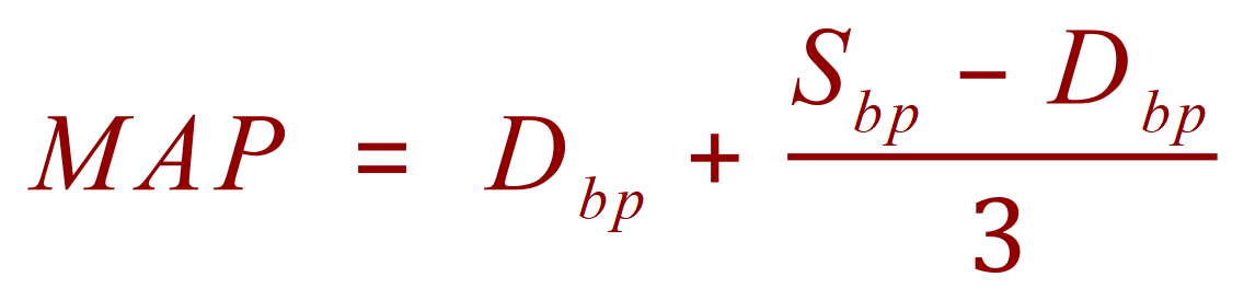
Where "MAP" refers to the mean arterial pressure and Dbp and Sbp refer to the diastolic and systolic pressures respectively.
Example:
A person, during a BP test records a systolic pressure of 145 mmHg and a diastolic pressure of 87 mmHg, what is the mean arterial pressure?

Questions that you will face at GCSE level usually involve rates of blood flow, and are fairly straightforward to calculate. At this level we consider these factors:
- STROKE VOLUME - The amount of blood pushed out from the heart through the left ventricle and into the aorta during each beat.
- HEART RATE - The number of beats that your heart makes per minute.
- CARDIAC OUTPUT - The product of the SV and HR as cubic centimetres per minute.

As is usually the case volume is measured in cubic centimetres.
Example:
The stroke volume of a person's left ventricle is taken as 70 cm³. During exercise the heart rate of the person increased to 125 bpm for a sustained period. What would be the Cardiac Output for one minute at this rate and volume?

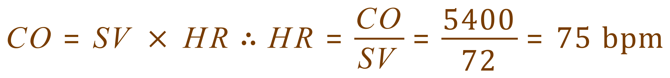
Example:
If the SV is 72 cm3 and the CO is 5400 cm3/min, what is the HR?