Microscopy
Microscopy is the study of very small objects using an instrument called a microscope. Generally there are two types of microscope, the light microscope which is the sort that you will use at school and the electron microscope which is used in industrial and high-level scientific investigation work. Of the two, you would naturally expect the electron microscope to be the more powerful and this is the case. A reasonably priced light microscope might cost you a couple of hundred pounds without electron microscopes are tens or even hundreds of thousands of pounds.
One of the required practical is in the biology specification, a practical investigation into the creation and examination of onion cells. You will be expected to be able to create a slide, stain the sample appropriately and obtain a good image from which you will hand draw what you can see. If this is done correctly you will able to see the cell walls and nuclei of the individual onion cells. The only real mathematics that comes into this is in calculating actual sizes and magnified sizes given the magnifications involved.
A typical light microscope will magnify from about 40 to 2500 times. Facilities will exist on most microscopes and slides to obtain a measurement of the image that you are observing, but of course it will not be the actual size of the specimen. The size of the specimen will depend on the magnification being used.
Q. A magnified image is 2 mm wide but you know that the actual specimen is 0.02 mm wide, given this information calculate the magnification on the microscope?
A. The magnification is given by the following quite simple equation:
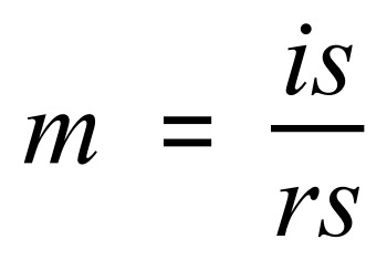
Where 'm' is the magnification being used, 'is' is the image size (as seen down the microscope) and 'rs' is the real size of the specimen.
We are giving the image size of 2 mm and the real size of the sample as 0.02 mm, we therefore work out the magnification thus:
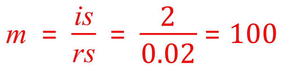
Our image is therefore created using a microscope with a magnification setting of 100x
Examiners like to have a little bit of fun every now and then, and so what they might do is give you the magnification and one of the measurements and ask you to transpose the equation in terms of the unknown one and then work it out. Let's now take a look at such an example:
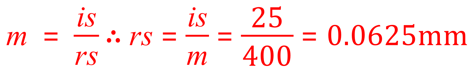
Q. A specimen viewed under a microscope gives an image size of 25 mm. The image is viewed at a magnification of 400x. What is the actual size of the specimen in millimetres?
A. You are given the magnification and the image size, so you need to transpose the above equation in terms of the unknown variable (the real size)
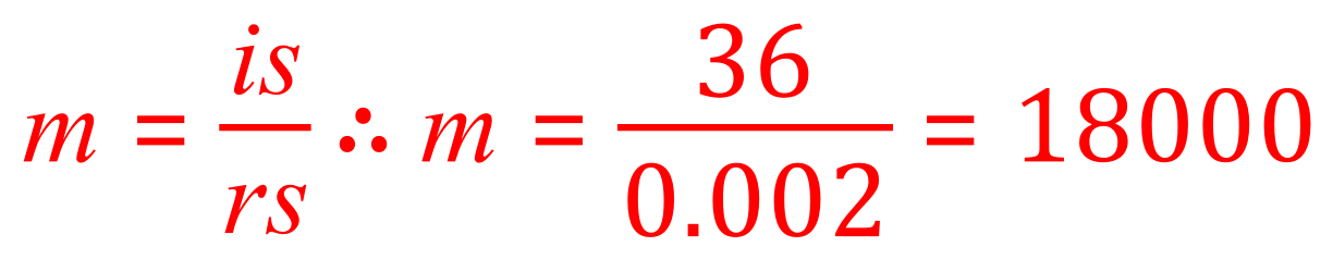
Q. A bacterial cell is 0.002 mm long, but under a microscope the magnified image measures 36 mm long. What is the magnification being used?
A. This is once more a simple "plug in" of the values given. You are given the image size 36 mm and you are given the real size which is 0.002 mm. Simply dividing one by the other will give you the magnification.
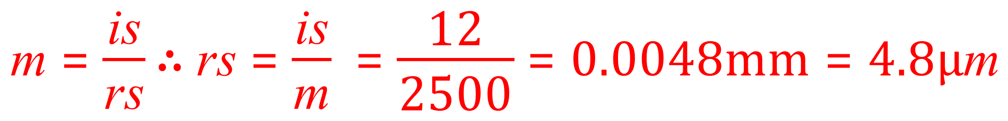
Q. A specimen is being observed under a light microscope at a magnification of 2500x. The magnified image is 12 mm wide. Calculate the actual size of the specimen and state your answer in micrometres (um).
A. This is a two-part question effectively, you need to be able to transpose the equation so that the actual width/size of the image is the subject and also convert your answer into micrometres. You may or may not be given the conversion factor between millimetres and micrometres, if this isn't the case you will need to know that 1mm = 1000 µM
First of all, let's rearrange the equation in terms of the unknown: